
Lung diseases are difficult to study because the alveoli constantly expand and contract, and this motion influences how lung cells grow, function, and respond to inflammation. Conventional cell‑culture systems cannot reproduce this movement, and even lung‑on‑a‑chip devices often rely on synthetic materials that fail to mimic real tissue. Researchers at the Pohang University of Science and Technology (POSTECH) in Korea developed an ultrathin artificial lung inspired by the stability and flexibility of soap bubbles, enabling realistic alveolar motion and allowing scientists to observe how lung cells behave under dynamic conditions.
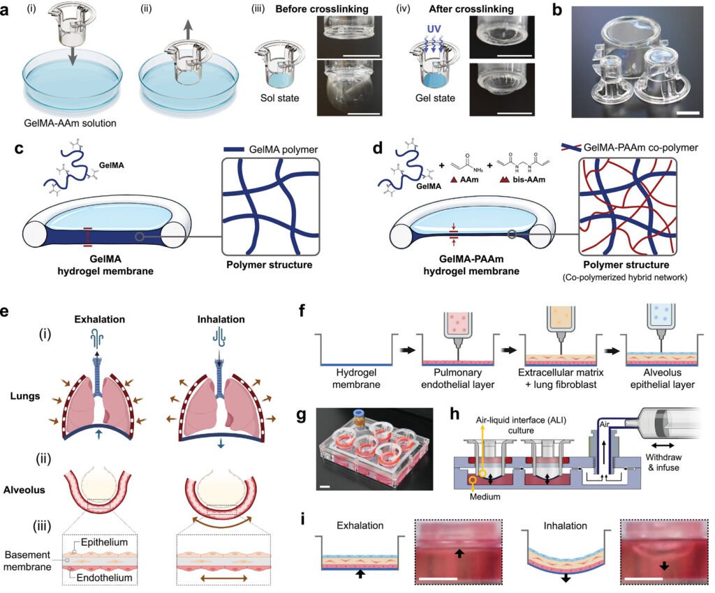
The team drew inspiration from the thin film of a soap bubble, which forms naturally, moves freely, and resists bursting. They applied this principle to a composite hydrogel made by mixing polymers and monomers. By dipping a mold into the hydrogel solution and withdrawing it, they produced an extremely thin membrane inside the mold. This membrane was soft and stretchable like native alveoli but strong enough to withstand hundreds of thousands of repeated motions. The artificial lung operated stably through approximately 240,000 breaths, demonstrating durability suitable for extended experiments.
To replicate real breathing, the researchers built a “breathing actuation system” that generates pressure changes similar to those created by the diaphragm. When the pressure beneath the ultrathin membrane fell, the membrane expanded; when the pressure rose, it contracted. This reproduced the inhalation and exhalation cycle and transmitted motion directly to the lung cells cultured inside the device.
The interior of the artificial lung was engineered to resemble the layered structure of alveoli. Using 3D bioprinting, the team stacked a vascular cell layer, a basement membrane layer, and an epithelial cell layer. Within this environment, the breathing motion influenced cell behavior, and the artificial lung reproduced responses similar to those seen in real tissue. Influenza virus infection experiments revealed that inflammatory and antiviral responses differed depending on respiratory motion, highlighting the importance of studying lung biology under dynamic conditions.
The researchers emphasized that this system enables drug‑response testing in a setting far closer to the human lung than traditional cell culture or animal models. Because the artificial lung allows the rate and depth of breathing to be freely adjusted, it can model both healthy and diseased states. They noted that the technology opens new possibilities for investigating lung disease mechanisms and evaluating therapeutics in a controlled, physiologically relevant environment.
Article from POSTECH: Develops a 'Breathing Lung' Inspired by Soap Bubbles
Abstract in Advanced Materials: Ultrathin Hydrogel Membranes Inspired by Soap Films Enable Physiologically Relevant Breathing Lung Models
