
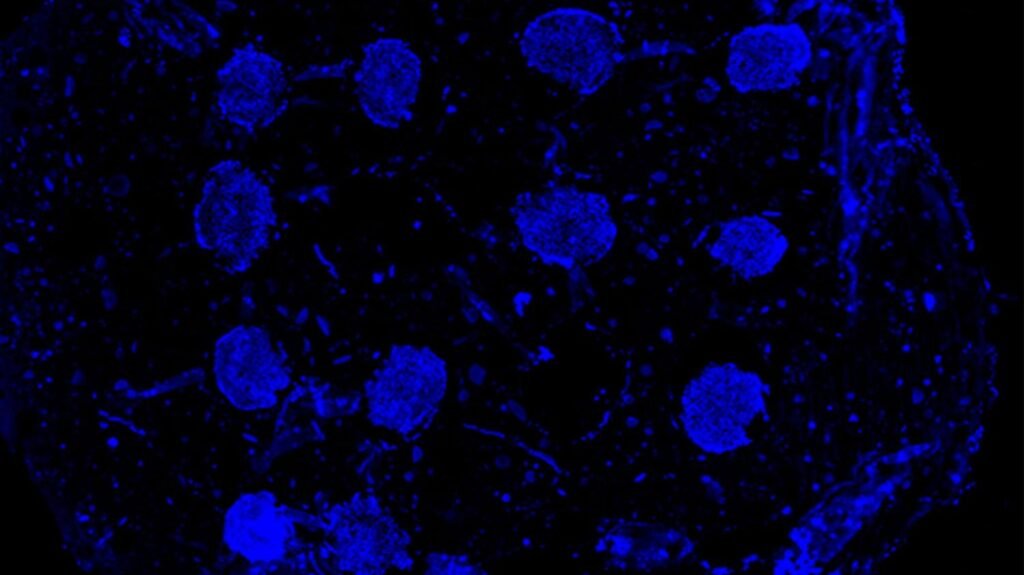
Severe bone injuries caused by trauma, cancer, or infection often fail to heal because engineered bone tissue struggles to form the blood vessels needed to deliver oxygen and nutrients. Without vascularization, even advanced bioprinted constructs cannot mature or integrate with the body. Researchers at Pennsylvania State University developed a strategy that programs living stem‑cell spheroids with genetic switches to promote both bone formation and blood‑vessel growth, addressing one of the central challenges in bone tissue engineering.
The team began with commercially sourced, undifferentiated stem cells and introduced specific strands of genetic information to guide their behavior. By layering these cells into spheroids using bioprinting, the researchers created compact clusters capable of supporting bone regeneration. The spheroids were positioned inside a microgel scaffold using a specialized 3D‑printing technique, allowing the cells to mature within a supportive environment. Experiments conducted in the lab and in mouse models showed that these engineered spheroids not only improved bone healing but also facilitated the formation of new blood vessels within regenerated tissue.
Bioprinting typically relies on hydrogels as scaffolds, which act as three‑dimensional matrices for cell growth. However, generating vascular networks inside bioprinted bone has historically been difficult. The Penn State team addressed this by using genetic programming to differentiate cells into distinct functional groups. Some spheroids were optimized for osteogenic activity, while others were programmed to support vascularization. This dual‑function approach allowed the researchers to build cellular networks with coordinated roles, a requirement for reconstructing complex tissues.
The genetic switches used in the study involved microRNA molecules that regulate gene activity. By influencing how cells mature and interact, these microRNAs helped direct spheroids toward bone‑forming or vessel‑forming pathways. The researchers verified that the spheroids maintained high viability and supported active cell behavior within the microgel matrix. In mouse studies, constructs containing genetically programmed spheroids improved bone regeneration and promoted the development of vessel‑like structures compared with controls.
The work establishes a potential framework for treating severe bone loss by enabling engineered tissues to develop both mineralized structure and vascular networks. The researchers noted that bioprinted spheroids could also serve as biological models for drug testing, but their primary focus is regenerative medicine. By demonstrating that genetic programming can coordinate multiple cell functions within bioprinted constructs, the Penn State team laid the groundwork for future therapies that rebuild bone tissue more effectively and reliably.
Article from Penn State: Genetic ‘switches’ could program 3D-printed bone tissue for blood vessel growth
Abstract in Chemical Engineering Journal: Bioprinting of miRNA-induced spheroids for vascularized, heterocellular bone regeneration
