
Heart disease remains difficult to study because conventional laboratory models cannot reproduce the complex motion, internal structures and disease mechanics of the human heart. Researchers at UNSW Sydney have developed a soft robotic model of the left side of the heart that mimics real cardiac behavior, including valve leakage and impaired relaxation, offering a controllable platform for studying disease and testing new medical devices. The fully synthetic system uses flexible materials, artificial muscles and detailed anatomical features to recreate how the heart contracts, twists and regulates blood flow.
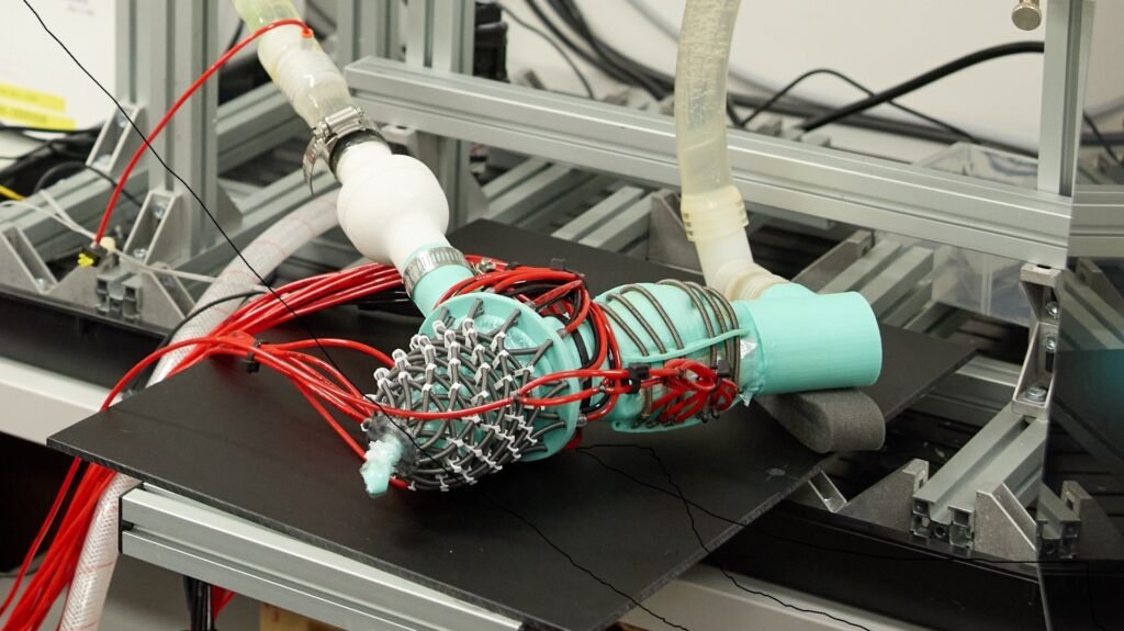
The model includes silicone membranes that form the internal chambers and soft robotic artificial muscles arranged to replicate the layered fiber architecture of human ventricular tissue. These muscles are powered by hydraulic pressure, allowing researchers to control contraction patterns and reproduce the natural twisting motion of the heart. The device also incorporates artificial papillary muscles and chordae tendineae, which support the mitral valve. By adjusting tension in these structures, the team can simulate conditions such as mitral valve prolapse and regurgitation, where blood flows backward instead of moving efficiently through the heart.
Ultrasound imaging and invasive pressure and flow measurements showed that the artificial heart behaves in ways closely aligned with human physiology. Healthy valve function produced normal pressure and flow patterns, while induced disease generated changes characteristic of valve dysfunction, including increased regurgitation and reduced outlet pressure. The model also reproduced imaging features seen in clinical echocardiography, such as human‑like valve leaflet motion and the formation of regurgitant jets. These capabilities allow researchers to study disease progression and evaluate how devices interact with moving cardiac structures.
The platform was used to test a soft robotic cardiac catheter, which successfully navigated within the beating model and detected contact with internal structures. Because the system offers repeatable control over heart function, the researchers believe it can reduce reliance on animal studies during early device development. They also demonstrated that the model can reproduce features of heart failure with preserved ejection fraction, including impaired relaxation and delayed filling that increase pressure inside the heart.
The team envisions future versions built from patient‑specific imaging data, enabling clinicians to evaluate devices and plan procedures using personalized heart models. Before clinical adoption, the system will require deeper validation against real patient data and further refinement of materials and control systems. The researchers view the current model as an enabling platform that can accelerate development of cardiac implants, improve surgical planning and support more effective treatment strategies for complex heart disease.
Take a look at this video showing more about the robotic heart:
Article from UNSW Sydney: Innovative soft robotic heart offers new way to study disease and test life-saving devices
Abstract in Nature Communications: Compliance modulation of a soft robotic atrioventricular model of heart failure with preserved ejection fraction