
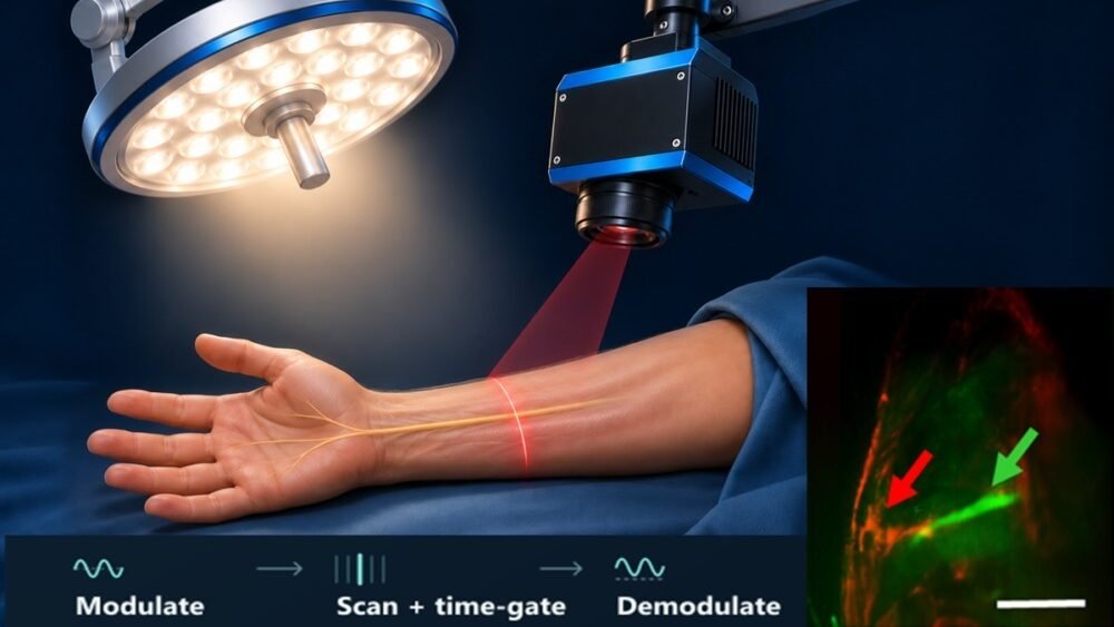
Accidentally damaging a nerve is a serious concern in many surgeries, and nerves can be hard to tell apart from surrounding tissue, especially in complex parts of the body. Fluorescence-guided surgery can make nerves easier to see by using fluorescent agents that selectively light up nerve tissue. The fluorescent signal, however, can be faint compared with the bright lights of an operating room, so surgeons may need to dim or turn off the room lights during imaging, which interrupts the normal flow of surgery. Researchers at Physical Sciences Inc. have developed an imaging system that separates the glow of labeled nerves from the much stronger background light, allowing nerves to be seen clearly under standard operating-room lighting.
The challenge is greatest for fluorescence at visible wavelengths, where light from surgical lamps can reach the camera along with the fluorescent signal, reducing contrast and potentially hiding the structures surgeons want to see. Rather than simply trying to block this unwanted light, the research team gave the fluorescence a recognizable pattern in both space and time that sets it apart from ordinary lighting.
The system uses line-scan imaging, collecting the fluorescent signal from one narrow region at a time. This encodes the fluorescence into a specific spatial pattern and limits its detection to a short window of time. Image processing then pulls out signals that match this pattern while suppressing background light, which stays relatively steady over time and even across the field of view.
Early studies showed that the imager could detect nerves despite strong ambient lighting. The research team then expanded testing from live animal models to nerve samples removed from humans and to amputated human limbs supplied with circulating fluid, which more closely resemble the conditions of human surgery. Across these experiments, the system produced high-contrast images of nerve tissue under normal operating-room lighting.
This matters for bringing fluorescence-guided nerve imaging into routine surgical practice, since a technology that forces surgical teams to repeatedly change their working conditions can be hard to adopt. Further clinical evaluation will be needed to see how the technique performs during human surgery. By adapting the imaging to the operating room rather than requiring the operating room to adapt to the imaging, the approach could make fluorescence-based nerve visualization easier to fit into surgical workflows.
Article in SPIE: Imaging nerves during surgery without turning down the lights
Abstract in Journal of Biomedical Optics: Spatio-temporally modulated fluorescence imager for intraoperative nerve identification
