
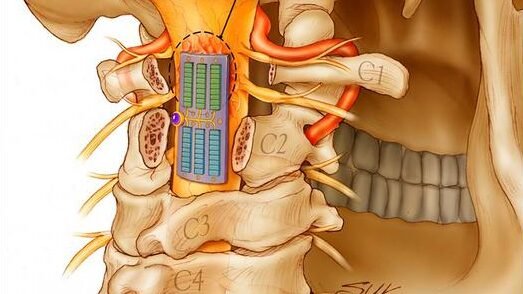
An estimated 18,000 spinal cord injuries occur in the United States each year, most often from vehicle accidents. After the surgery that typically follows such an injury, patients recover in the intensive care unit, where they remain at risk for serious complications such as stroke, blindness, or further loss of movement control. Getting a picture of what is happening at the injury site often means imaging that requires moving patients who cannot move on their own, and it captures only single moments in time. Engineers and surgeons at Johns Hopkins Medicine have now completed a first proof-of-concept study showing that a tiny ultrasound device can be temporarily implanted at the injury site and provide continuous feedback.
The device, called the “Multimodal Ultrasound System for Injury Care”, is about the size of a small bandage and can be placed over the spinal cord during the surgery that follows an injury. It is designed to act as a kind of mobile imaging scanner, so medical teams can check on the injury whenever they need to without moving the patient. The goal is to give clinicians better information about what is happening at and around the injury during recovery and to support their decisions about care.
The device combines two forms of ultrasound. Its diagnostic side delivers real-time information about blood flow and other changes around the injury. It can also help assess spinal fluid, excess fluid buildup, how a patient is responding to medication, and what is happening beyond the injured area, all of which can offer clues about how well the person is healing. This kind of immediate feedback in the days and weeks after surgery could help doctors personalize treatment. Its therapeutic side uses focused ultrasound, a more exploratory feature that is being studied for its potential to detect and respond when blood flow through the small vessels around the injury rises above or falls below acceptable levels.
The device received Breakthrough Device Designation from the U.S. Food and Drug Administration in 2023. In the new study, conducted in animals, the team showed that the device could be temporarily implanted and provide real-time imaging and blood flow information around the site where it was placed. The researchers found the approach to be feasible, safe, and potentially useful.
The findings mark an early milestone in a multistage research effort. Through further preclinical studies, the research team will continue exploring how the device can be optimized to track blood flow, identify changes in the tissues and spine, and monitor other activity near the injury. The team designed the device to help people of many ages and backgrounds, from soldiers wounded on the battlefield to children and adults injured in car crashes, falls, or sports. The researchers believe that fine-tuning care in the critical period after surgery could improve patients’ health outcomes months and years later.
Here’s a short video from Johns Hopkins explaining the device:
EurekAlert! Press Release: Researchers move closer to advancing spinal cord injury care
Abstract in Science Advances: Ultrasound neural interface enabling acute spinal cord monitoring and blood flow modulation
