
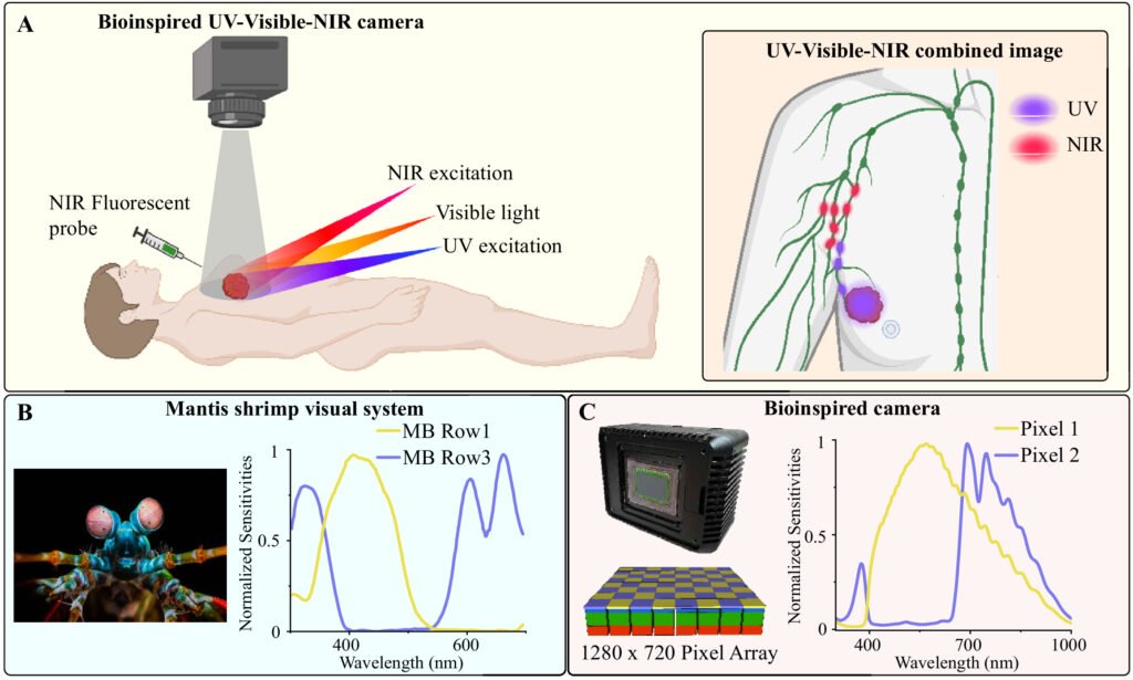
Cancer surgery often hinges on a difficult decision. Surgeons must determine which lymph nodes are likely to contain cancer and which should be preserved to avoid long term complications. Existing imaging tools can show where lymph fluid drains, but they cannot reliably indicate whether a specific lymph node is cancer linked during the operation. This gap forces surgeons to rely on experience and partial information at a moment when accuracy matters most. Researchers at the University of Illinois Urbana-Champaign have now introduced a compact imaging system that brings a new level of clarity to this decision by capturing ultraviolet, visible, and near infrared information on a single chip. The goal is to give surgeons real time insight into both the location of lymph nodes and the biochemical signatures that may signal cancer involvement.
The device draws inspiration from the mantis shrimp, a marine animal known for its extraordinary ability to detect many wavelengths of light with high precision. This biological model guided the design of a sensor that separates multiple wavelengths at the pixel level. The system uses near infrared light to locate lymph nodes and ultraviolet light to detect biochemical features that correlate with cancer. Because all wavelengths are captured on the same chip, the images remain perfectly aligned. This solves a long standing problem in surgical imaging, where different modalities often produce images that do not match up cleanly.
Early tests with breast cancer tissue showed that the camera can identify lymph nodes and highlight suspicious biochemical patterns without the need for dyes or contrast agents. The ability to perform label free assessment during surgery could reduce the number of lymph nodes removed unnecessarily. It could also help surgeons avoid missing nodes that are more likely to contain cancer. The compact size of the device makes it easier to integrate into operating rooms without disrupting existing workflows.
The researchers believe this technology could improve outcomes for patients by supporting more precise surgical decisions. It may also reduce the need for repeat procedures and lower the risk of complications such as lymphedema. Beyond breast cancer, the approach could be applied to other cancers where lymph node status guides treatment. The combination of multispectral imaging and perfect pixel level alignment opens the door to new forms of real time tissue assessment that fit naturally into the pace of surgery.