
Cancer drugs often fail because they do not reach the right part of the cell, yet scientists have had no reliable way to track where a drug accumulates inside a living cell. Existing methods usually require killing the cell first, which destroys the very information researchers need. Scientists at the University of Surrey and King’s College London have developed a new analytical technique that solves this long‑standing problem by mapping drug uptake inside individual living cells and even inside their internal structures.
The method was created to study targeted radionuclide therapy, a cancer treatment that attaches a radioactive particle to a molecule designed to seek out tumor cells. The location of the drug inside the cell is critical because a drug that reaches the nucleus can damage DNA, while a drug that remains elsewhere may have little effect. Until now, researchers could not measure this distribution in living cells.
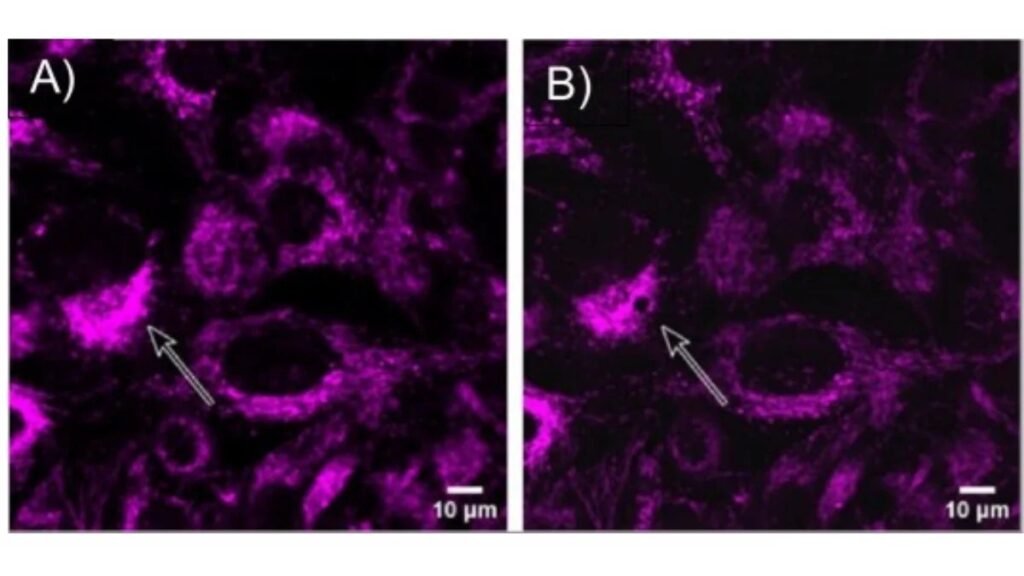
The team combined two specialized technologies into a single workflow. First, they used the SEISMIC facility at King’s College London, which employs tiny glass capillary tips to extract material from living cells without killing them. The tips are extremely small, about ten micrometers wide for whole cells and three micrometers for subcellular structures such as mitochondria. Second, they used laser ablation inductively coupled plasma mass spectrometry at the University of Surrey to detect trace amounts of metal inside the sampled material. This combination allowed the researchers to measure thallium, used as a stand‑in for a radioactive cancer drug, inside both whole cells and mitochondria. This level of subcellular mapping in living cells had not been achieved before.
The researchers demonstrated that thallium could be detected at extremely low levels inside individual pancreatic cancer cells and inside mitochondria‑enriched samples taken from those cells. This provides a new way to determine whether a drug reaches the structures it is meant to target.
By enabling precise, real‑time mapping of drug distribution inside living cells, the technique could help scientists design more effective cancer therapies. It offers a clearer view of how drugs behave once they enter a cell and could guide the development of treatments that deliver their active components exactly where they are needed.
Article from the University of Surrey: New technique maps cancer drug uptake inside living cells
Abstract in Spectrochimica Acta Part B: Atomic Spectroscopy: Subcellular capillary sampling coupled to laser ablation – Inductively coupled plasma – Mass spectrometry (LA-ICP-MS) allows targeted analysis of thallium in a radiopharmaceutical model