
A new imaging breakthrough from Northwestern University could reshape nuclear medicine by making scans faster, clearer, and more affordable. Researchers have built the first gamma-ray detector using perovskite crystals, a material best known for its role in solar energy. This detector captures individual gamma rays with record-setting precision, offering a powerful alternative to current technologies used in SPECT imaging.
SPECT, or single-photon emission computed tomography, is a common diagnostic tool that tracks blood flow, heart function, and hidden diseases using radiotracers. These tracers emit gamma rays that pass through the body and are collected by detectors to form detailed 3D images. However, existing detectors made from cadmium zinc telluride (CZT) or sodium iodide (NaI) are either prohibitively expensive or produce blurry results.
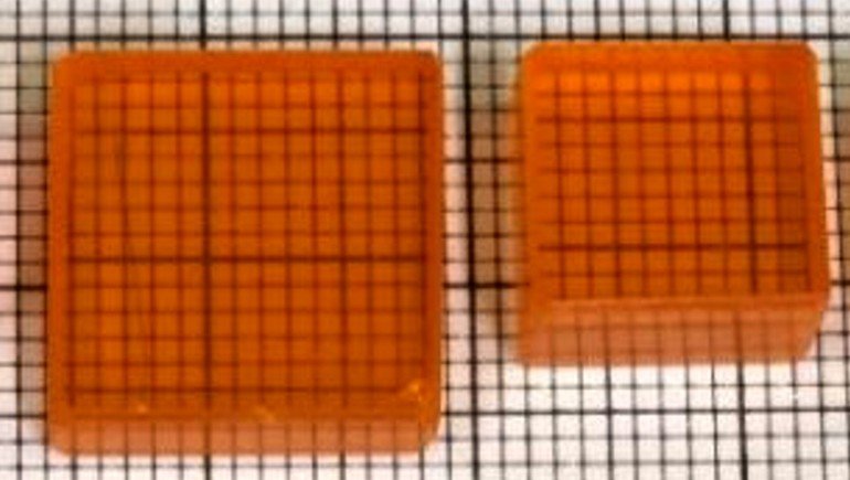
Perovskite crystals solve both problems. They are easier to grow, less fragile, and significantly cheaper than CZT, while offering superior resolution compared to NaI. The new detector uses a pixelated architecture similar to a smartphone camera, allowing it to distinguish fine details and detect faint signals from widely used radiotracers like technetium-99m.
In lab tests, the perovskite detector achieved the highest energy resolution reported to date and successfully imaged tiny radioactive sources spaced just millimeters apart. It remained stable throughout the experiments, collecting nearly all of the tracer’s signal without distortion. These improvements could allow for shorter scan times and lower radiation doses for patients.
Article from Northwestern University: First ‘perovskite camera’ can see inside the human body
Abstract in Nature Communications: Single photon γ-ray imaging with high energy and spatial resolution perovskite semiconductor for nuclear medicine