
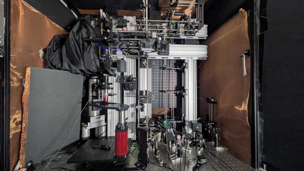
MIT researchers have developed a pioneering imaging system that enables unprecedented views into living brain tissue, reaching depths previously unattainable while maintaining single-cell resolution. Remarkably, this technique works without the need for chemical labels or genetic modifications.
The system relies on detecting NAD(P)H, a molecule closely tied to cellular metabolism and neuronal activity. Instead of traditional near-UV light, the microscope uses ultra-short pulses of light at three times the standard absorption wavelength—a method known as three-photon excitation. This longer wavelength penetrates deeper into tissue with minimal scattering, much like fog lamps illuminating through mist.
To capture the resulting signal, the team employs photoacoustic detection. When the targeted molecules absorb the light, they undergo tiny thermal expansions that generate sound waves. These waves are picked up by an ultrasound microphone and translated into high-resolution images.
The researchers successfully imaged a 1.1 mm thick human-derived cerebral organoid and a 0.7 mm thick slice of mouse brain—depths more than five times greater than what current NAD(P)H imaging technologies can achieve in dense brain tissue.
Article from MIT: MIT imaging tech promises deepest looks yet into living brain tissue at single-cell resolution
Abstract in Light: Science & Applications: Multi-photon, label-free photoacoustic and optical imaging of NADH in brain cells