
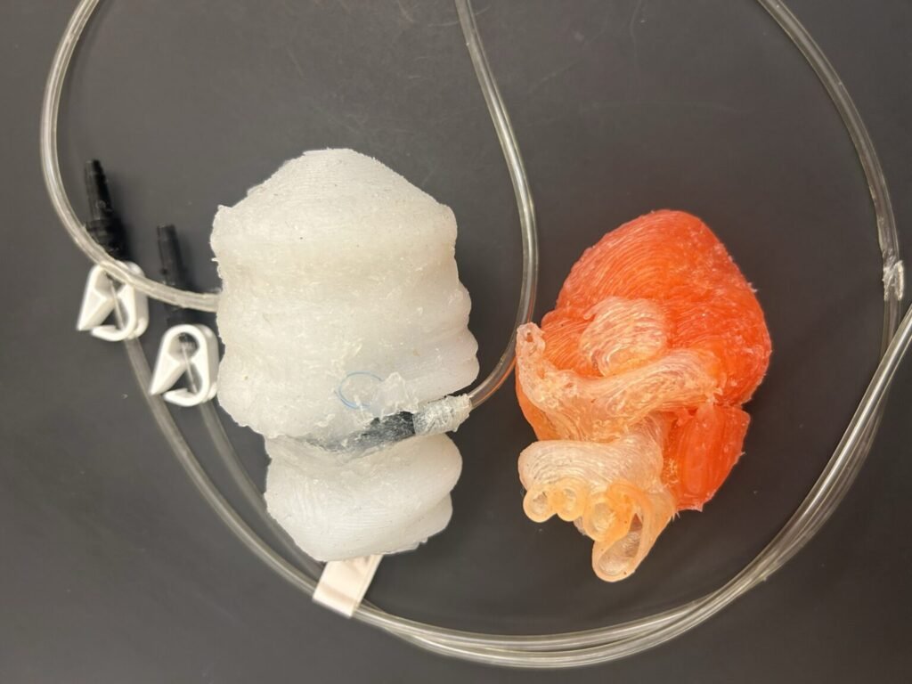
Researchers at Washington State University have created a 3D‑printed heart model that can beat, pump fluid, and mimic the mechanical behavior of real cardiac tissue, offering surgeons a more realistic way to practice complex procedures before entering the operating room. The project grew out of a need for training tools that better replicate the feel and movement of a living heart. Traditional silicone models are static and lack the dynamic qualities that make cardiac surgery so challenging. The WSU team set out to build a model that could reproduce the motion, pressure changes, and tactile feedback of a functioning human heart.
The researchers used a custom 3D‑printing process to fabricate soft, flexible structures that match the elasticity of cardiac tissue. They then integrated pneumatic systems that rhythmically inflate and deflate the chambers, creating a lifelike beating motion. The model can pump fluid through its chambers, allowing surgeons to practice procedures that involve cutting, suturing, or navigating instruments inside a moving, fluid‑filled environment. This capability is especially important for training on delicate repairs where timing and precision are critical.
A key advantage of the system is its customizability. The team can print hearts that replicate specific patient anatomies, including congenital defects or unusual structural variations. This means surgeons could rehearse a procedure on a personalized model before performing it on the patient, improving preparedness and reducing risk. The researchers emphasize that the materials used in the model provide realistic resistance and texture, giving trainees a more accurate sense of how tissue behaves during surgery.
The project also aims to make advanced surgical training more accessible. Because the models can be printed relatively quickly and at lower cost than many commercial simulators, they could be used in medical schools, residency programs, and hospitals that lack access to high‑end training equipment. The team is continuing to refine the technology, exploring ways to incorporate sensors that track surgical performance or provide feedback on technique.
By combining soft 3D printing with engineered motion and fluid dynamics, the WSU researchers have created a beating heart model that brings surgical practice closer to real clinical conditions. The technology offers a promising path toward safer, more effective training for cardiac surgeons and could help improve outcomes for patients undergoing complex heart procedures.
Article from WSU: Researchers develop beating, 3D‑printed heart model for surgical practice
Abstract in Advanced Materials Technologies: 3D-Printed Dynamic Heart Model With Left-Side Anatomy and Integrated Sensor for Edge-to-Edge Repair and Regurgitation Reduction