
A new development from MIT introduces a portable ultrasound sensor designed to make breast cancer screening more frequent, accessible, and timely for people at elevated risk. The research team created a compact imaging system that can be used at home or in a doctor’s office, addressing a long‑standing challenge in early detection. Many high‑risk individuals struggle to access regular imaging due to cost, scheduling barriers, or limited availability of specialized equipment. The MIT system aims to close that gap by offering a low cost, easy to use alternative that still provides clinically meaningful imaging.
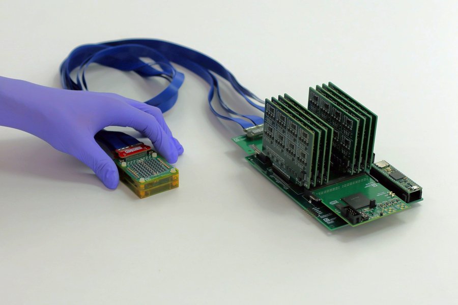
The device consists of a small ultrasound probe connected to an acquisition and processing module that is slightly larger than a smartphone. The probe itself is about the size of a deck of cards and contains an ultrasound array arranged in an open square configuration. This geometry allows the system to capture three dimensional images of breast tissue, giving users and clinicians a more complete view of potential abnormalities. The researchers emphasize that the system’s compact design does not compromise imaging capability. Instead, it enables real time 3D scanning that can be performed more frequently than traditional clinical ultrasounds.
One of the central motivations behind the project is the importance of early detection. Breast cancer outcomes improve significantly when tumors are identified at an early stage, yet many people only receive annual or biennial imaging. The MIT team designed the portable sensor to support more regular monitoring, particularly for individuals with dense breast tissue or genetic risk factors. Because the system is relatively inexpensive to manufacture, the researchers believe it could expand access in underserved regions and rural communities where imaging facilities are limited.
The device can connect to a laptop for real time visualization, allowing users or clinicians to view 3D images immediately. The team envisions future versions that integrate with mobile devices to further streamline use. The researchers also highlight the potential for personalized screening schedules, where high risk individuals could perform scans at home between clinical visits. This approach could help identify suspicious changes earlier and reduce delays in diagnosis.
By miniaturizing ultrasound hardware and pairing it with an accessible processing module, the MIT team has created a platform that could shift breast cancer screening toward more frequent, user driven monitoring. The technology offers a practical path to earlier detection, especially for people who face barriers to traditional imaging, and reflects a broader movement toward portable, patient centered diagnostic tools.
Article from MIT: A portable ultrasound sensor may enable earlier detection of breast cancer
Abstract in Advanced Healthcare Materials: Real-Time 3D Ultrasound Imaging with an Ultra-Sparse, Low Power Architecture