
Glioblastoma is one of the most aggressive brain cancers, with most patients dying within two years and only about 10 percent surviving five years. Surgery cannot remove all tumor cells because they infiltrate brain tissue, and most chemotherapy drugs cannot cross the blood‑brain barrier. This makes monitoring treatment effectiveness especially difficult.
Researchers at Northwestern Medicine and the University of Michigan have shown that a simple blood draw can reveal whether chemotherapy is killing glioblastoma cells. In a clinical trial, Northwestern doctors used the SonoCloud‑9 ultrasound device, developed by Carthera in France, to temporarily open the blood‑brain barrier for about an hour. This allowed the chemotherapy drug paclitaxel to reach the tumor.
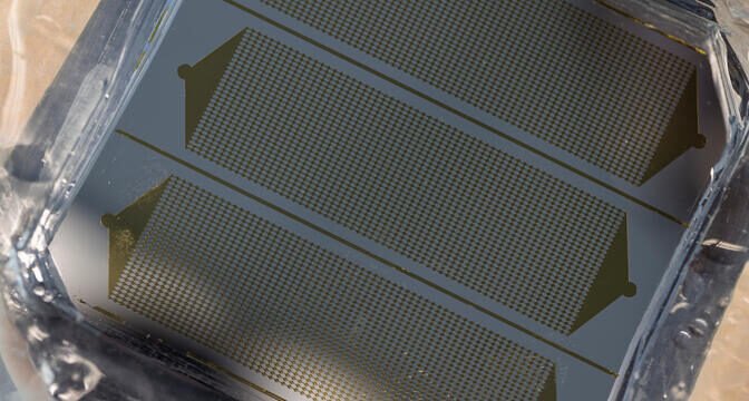
The Michigan team then applied their GlioExoChip, a diagnostic chip designed to capture extracellular vesicles and particles (EVPs) released by tumor cells into the blood. These vesicles carry genetic material and proteins from cancer cells. By isolating them with a lipid molecule found on their surface, the chip turns blood samples into liquid biopsies.
Counting EVPs before and after chemotherapy provided a clear signal. If the ratio of vesicles increased after treatment, it indicated that cancer cells were dying. If the ratio stayed flat or declined, the treatment was not effective. This method offers a minimally invasive way to monitor therapy response, avoiding the delays and misleading results that often come with MRI scans.
Researchers emphasized that patients could know after one dose whether a treatment is working, sparing them prolonged ineffective therapies and unnecessary side effects. They also explained that EVPs can be hijacked for disease progression but can also be leveraged to track treatment response.
Article from the University of Michigan: Blood analysis shows whether brain cancer treatment is working
Abstract in Nature Communications: Dynamic release of extracellular particles after opening of the blood-brain barrier predicts glioblastoma susceptibility to paclitaxel