
Strokes often begin with tiny blood clots forming inside damaged or irregular arteries, but it has been hard to watch those first moments in action. A University of Sydney team has built small, patient‑specific models of blood vessels that let researchers see how clots start and grow under lifelike conditions. They begin with medical scans from real patients and use those as blueprints to create miniature versions of the carotid artery, the vessel in the neck that supplies blood to the brain. These printed vessels are made directly on glass, which helps blood flow more naturally through them during tests. The goal is to understand which spots inside a vessel are most likely to spark platelets to clump together and form a clot.
Speed and realism are the big advantages. Traditional lab methods can take many hours to build and often do not capture the way blood actually moves, especially in people with thicker, more viscous blood due to heart disease. The new approach can produce a test‑ready vessel in about two hours and preserves fine details like dents and divots on the inner surface that are common in patients after vascular damage. When blood is run through these models, researchers can watch platelets move, stick, and cluster. They have seen that areas under high mechanical stress show much more platelet activity, which helps explain why some regions are more prone to clotting.
Because every person’s blood and vessel shape are different, printing a model from an individual’s scan opens the door to personalized testing. Imagine a workflow where a patient’s vessel is printed, their blood is tested in that model, and doctors get a readout of how likely clots are to form in that specific geometry. That kind of information could guide decisions on blood pressure targets, medication choices, and timing of interventions. It could also help doctors understand why two patients with similar narrowing of an artery face different levels of risk.
The team is also exploring how to pair these physical models with computer predictions. By analyzing blood flow maps, surface features inside the vessel, and the motion of platelets, software could estimate clot risk before symptoms appear. In practical terms, a patient might have a scan on day one, receive a printed vessel model within hours, and get a tailored risk assessment shortly after. This kind of precision could help prevent strokes by identifying problems early and focusing care on the highest‑risk spots inside the artery.
Beyond stroke, the same platform could shed light on clotting in other conditions, such as peripheral artery disease or areas near stents where blood flow can change. Replacing animal tests with patient‑specific models makes the research more humane and more relevant to real human biology. For patients, it means clearer answers and potentially faster, more personalized care. For clinicians and engineers, it offers a reliable way to watch clot formation in real time and to test new treatments on models that closely mirror the vessels they see in practice. By making the earliest steps of clotting visible and measurable, these printed blood vessels bring stroke prevention closer to everyday clinical use.
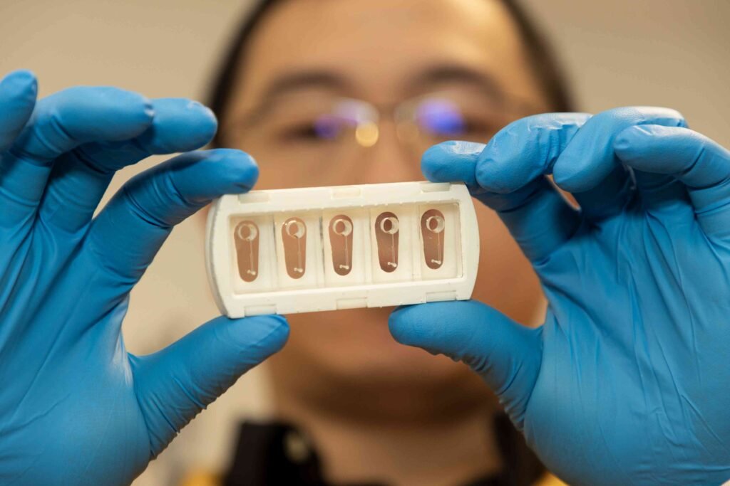
Article from the University of Sydney: 'Artery on a chip': 3D printed blood vessels could unravel secrets of strokes
Abstract in Advanced Materials: Rapid Glass-Substrate Digital Light 3D Printing Enables Anatomically Accurate Stroke Patient-Specific Carotid Artery-on-Chips for Personalized Thrombosis Investigation