
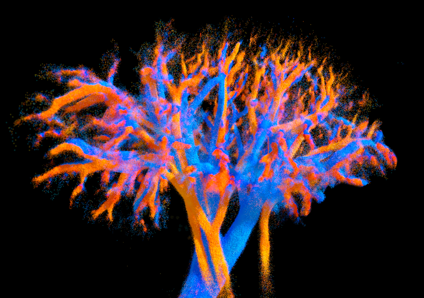
A new ultrasound imaging system developed by researchers at Inserm’s Physics for Medicine Institute in Paris is redefining how clinicians visualize blood flow across entire organs. The probe captures four-dimensional imaging—three spatial dimensions plus time—allowing dynamic, high-resolution views of vascular networks in organs such as the heart, liver, and kidneys. This technology offers a rare combination of full-organ coverage and micrometer-level precision, which has long been a trade-off in conventional ultrasound systems.
The device uses 252 acoustic lenses arranged across a flat 8 by 10 centimeter array. This multi-lens configuration enables simultaneous imaging of volumes up to 120 by 100 by 82 millimeters. Unlike traditional ultrasound systems that scan sequentially or sacrifice resolution for field of view, this design allows real-time visualization of blood flow patterns, including velocity and direction. Researchers demonstrated the system’s ability to distinguish arteries from veins and track vascular dynamics in preclinical animal models.
The probe is compatible with existing ultrasound platforms and compact enough for clinical use. Its ability to visualize microcirculation and vascular behavior across entire organs could improve diagnosis and monitoring of cardiovascular disease, cancer, and transplant viability. The team envisions applications in surgical guidance, noninvasive vascular assessment, and early detection of organ dysfunction.
Here’s a YouTube short illustrating the new imaging system:
Article from Inserm: Researchers develop an ultrasound probe capable of imaging an entire organ in 4D
Abstract in Nature Communications: Multi-lens ultrasound arrays enable large scale three-dimensional micro-vascularization characterization over whole organs