
Studying Alzheimer’s disease in living, active animals has long been a challenge due to limitations in imaging technology. Researchers at the University of Strathclyde and the Italian Institute of Technology have now developed a fiber-optic method that allows scientists to track amyloid-beta plaques in the brains of freely moving mice. This breakthrough offers a more realistic view of how the disease progresses and how plaques behave in natural conditions, rather than under anesthesia or in restrained environments.
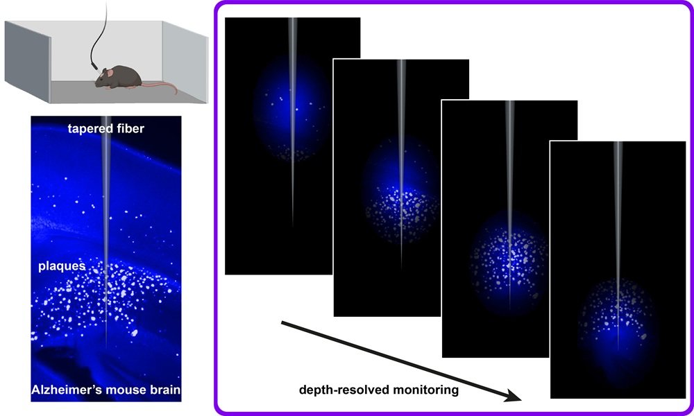
The technique uses a specialized fiber-optic probe that emits and collects light to detect fluorescent markers bound to amyloid-beta plaques. These markers glow when exposed to specific wavelengths, allowing researchers to monitor plaque formation and distribution in real time. The probe is small and flexible enough to be implanted in the brain without interfering with the animal’s movement, making it possible to observe disease dynamics during normal behavior.
One of the key advantages of this method is its ability to capture data continuously over extended periods. Researchers can now study how plaques evolve in response to environmental factors, physical activity, or therapeutic interventions. This opens the door to more accurate testing of potential treatments and a better understanding of how lifestyle or drug regimens might influence disease progression.
The fiber-optic system also enables high-resolution imaging without the need for bulky equipment or invasive procedures. It complements existing imaging techniques like two-photon microscopy but offers greater flexibility and accessibility for long-term studies. The researchers believe this approach could eventually be adapted for use in other neurological conditions that involve protein aggregation or inflammation.
Article from SPIE: New fiber-optic method tracks Alzheimer’s plaques in active mice
Abstract in Neurophotonics: Depth-resolved fiber photometry of amyloid plaque signals in freely behaving Alzheimer’s disease mice