
Researchers at Radboud University Medical Center have developed a powerful new tool that combines artificial intelligence with a miniature imaging technique to predict which patients are at risk of future heart attacks. The system uses optical coherence tomography, a catheter-based camera that captures detailed images inside coronary arteries, and AI to identify vulnerable plaques that could rupture and block blood flow.
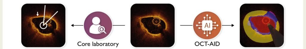
In a study involving 438 patients, the AI system matched and even outperformed expert labs in identifying high-risk plaques. It also proved better at predicting which patients would suffer a recurrent heart attack or die within two years. This is a major advance, as current methods often miss subtle signs of plaque instability, and interpreting OCT scans manually is time-consuming and expensive.
OCT is already used to guide stent placement during heart procedures, but its full diagnostic potential has been limited by the sheer volume of data it produces. Hundreds of images are generated per scan, making full-artery analysis impractical without automation. The AI streamlines this process, analyzing entire arteries quickly and consistently. This makes it feasible to use OCT not just for treatment but for proactive risk assessment.
The technology could lead to more personalized care. Instead of treating all patients with the same medications or waiting for symptoms to worsen, doctors could identify those at highest risk and intervene earlier through preventive stenting, lifestyle changes, or targeted therapies.
The research team, led by Dr. Jolanda Wentzel and Dr. Evelyn Regar, believes this approach could transform how cardiologists manage coronary artery disease. By focusing on the biology of individual plaques rather than general risk factors, the system offers a more precise way to prevent heart attacks before they happen.
Press Release: Combination of mini-camera and AI predicts recurrent heart attack
Abstract in the European Heart Journal: Artificial intelligence-based identification of thin-cap fibroatheromas and clinical outcomes: the PECTUS-AI study