
Researchers at the University of Colorado Boulder have developed a next-generation bioimaging device that could dramatically improve how doctors detect and monitor conditions affecting the eyes and heart. Unlike traditional imaging systems that rely on mechanical components like spinning mirrors, this new device operates entirely without moving parts—making it more compact, energy-efficient, and durable.
The technology builds on optical coherence tomography (OCT), a widely used method in eye clinics that uses light waves to produce high-resolution, cross-sectional images of the retina. OCT is essential for diagnosing and tracking diseases such as macular degeneration and glaucoma. However, current OCT systems are bulky and mechanically complex, which limits their portability and increases the risk of mechanical failure over time.
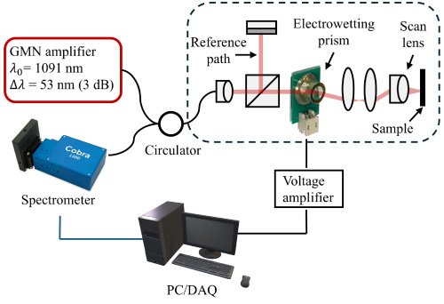
To address these limitations, the Colorado team created a nonmechanical imaging system based on a process called electrowetting. This technique manipulates the surface shape of a liquid using electrical signals, allowing the device to perform optical functions without physically moving any components. The result is a lightweight, low-power imaging tool that could be used not only in eye exams but also for in-vivo imaging inside the body—including the heart and other organs.
Lead researcher Samuel Gilinsky, a recent PhD graduate in electrical engineering, emphasized the potential of the device for retinal imaging and broader biomedical applications. By eliminating mechanical parts, the system reduces wear and tear, improves reliability, and enhances safety for use in sensitive areas of the body. The device also consumes significantly less power than conventional OCT systems, making it ideal for portable or wearable formats.
To test the device’s capabilities, the researchers used zebrafish—a model organism with eye structures similar to humans. The team successfully captured detailed images of the cornea and iris, achieving resolution benchmarks of 10 microns axially and 5 microns laterally, both smaller than the width of a human hair. These results confirmed that the device could resolve fine biological structures with precision.
The research team included electrical engineering professors Juliet Gopinath and Shu-Wei Huang, mechanical engineering professor Victor Bright, PhD graduates Jan Bartos and Eduardo Miscles, and PhD student Jonathan Musgrave. Their work was published in Optics Express, a leading journal in photonics and imaging science.
Professor Gopinath noted that the device could help detect health conditions earlier and improve patient outcomes. Its compact design and nonmechanical operation make it well-suited for integration into wearable health monitors or portable diagnostic kits—potentially expanding access to advanced imaging in remote or underserved areas.
Article from the University of Colorado: New bioimaging device holds potential for eye and heart condition detection
Abstract in Optics Express: Nonmechanical spectral domain optical coherence tomography using an electrowetting beam-scanner