
Scientists from Helmholtz Munich, the Technical University of Munich, and the Medical University of Vienna have created a new endoscopy capsule that could dramatically improve early detection of esophageal cancer. This type of cancer is often deadly when diagnosed late, but survival rates soar when caught early. The new technology, called O2E, combines two advanced imaging techniques to reveal even the smallest changes in tissue and blood vessels.
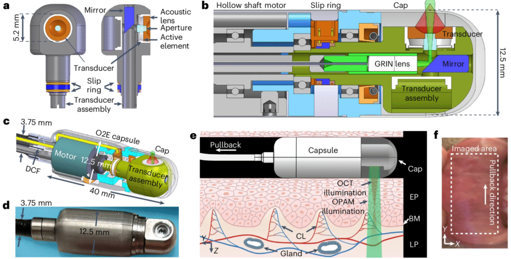
The capsule uses optical coherence tomography to map tissue structure and optoacoustic imaging to visualize blood flow by detecting sound waves generated by light pulses. Together, these methods produce detailed 3D images of the esophagus, allowing doctors to spot abnormalities that older tools might miss. The capsule scans in a full 360-degree sweep and has already shown success in identifying precancerous and cancerous changes in both animal models and human tissue samples.
Researchers are now working to refine the device for clinical use and plan to add even more imaging capabilities, such as confocal endomicroscopy, which can zoom in on individual cells. The goal is to reduce the need for multiple biopsies and speed up diagnosis.
Article from Helmholtz Munich: New Endoscopy Technology Enables Early Detection of Esophageal Cancer
Abstract in Nature Biomedical Engineering: Tethered optoacoustic and optical coherence tomography capsule endoscopy for label-free assessment of Barrett’s oesophageal neoplasia