
For the 300,000 Americans living with scleroderma—a rare autoimmune disease that causes tissue scarring in organs like the lungs, liver, and skin—treatment options remain limited and unpredictable. Now, researchers at Tufts University and Dartmouth’s Geisel School of Medicine have developed a 3D tissue model that replicates the complexity of fibrotic disease more accurately than traditional lab methods. This breakthrough could lead to faster, more personalized therapies for scleroderma and other conditions involving fibrosis.
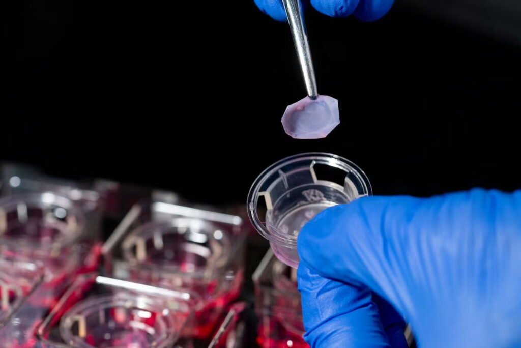
The model is built using skin and blood samples from scleroderma patients and includes two key immune cells—T cells and macrophages—alongside connective tissue cells. These immune cells are central to fibrosis, driving the overproduction of collagen that stiffens and damages tissue. Unlike conventional 2D cell cultures, which flatten and homogenize cell behavior, the 3D model preserves the natural diversity and architecture of living tissue. Under the microscope, the lab-grown skin is nearly indistinguishable from real skin.
This fidelity allows researchers to observe how fibrosis progresses differently in different individuals and to isolate single cells for gene expression analysis. By identifying which genes are active in specific cell types, scientists can better predict which treatments will work for which patients—whether they have localized skin thickening or multi-organ involvement.
The model also offers a scalable, ethical alternative to animal testing and donated tissue samples, which are often scarce and inconsistent. It can be adapted to study other fibrotic diseases, including pulmonary and myocardial fibrosis, and may help accelerate drug development by providing a more predictive platform for testing safety and efficacy.
Article from Tufts University: New 3D Tissue Model May Speed Better Therapies for Fibrosis
Abstract in Tissue Engineering Part C: Methods: T Cells Enhance Tissue Complexity and Function to Study Fibrosis in 3D Skin-Like Tissue Models