
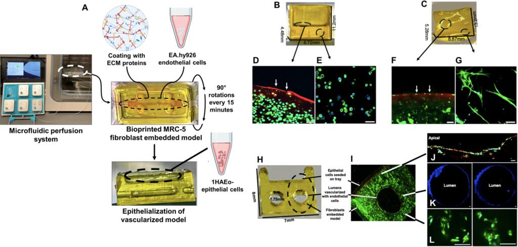
Researchers at UBC Okanagan have developed a 3D bioprinted model that closely mimics the complexity of living lung tissue, marking a major advance in how scientists study respiratory diseases and test new treatments. Led by Dr. Emmanuel Osei, the team used a specialized bioink composed of polymer-modified gelatin and polyethylene glycol diacrylate to print a hydrogel scaffold embedded with multiple cell types and vascular channels. This structure replicates the mechanical and biological properties of human airways, allowing researchers to observe cellular responses to disease triggers in a more physiologically relevant environment.
The model was validated by exposing it to cigarette smoke extract, which triggered inflammatory responses similar to those seen in real lung tissue—such as elevated cytokine levels. This ability to simulate disease progression makes the platform ideal for studying conditions like asthma, COPD, idiopathic pulmonary fibrosis, and lung cancer. It also enables researchers to test drug candidates without relying on donated tissue samples, which are often scarce and inconsistent.
Importantly, the bioprinted lung tissue can be customized with patient-derived cells, opening the door to personalized medicine and more targeted therapies. The reproducibility and modularity of the system make it a powerful tool for both academic research and pharmaceutical development. Supported by Mitacs and Providence Health Care, the study positions UBCO’s team to collaborate with biotech companies and immunology researchers to further refine the model and explore its use in regenerative medicine.
Article from UBC Okanagan: UBCO researchers create 3D-printed living lung tissue
Abstract from Biotechnology and Bioengineering: Establishing a 3D Vascularized Tri-Culture Model of the Human Airways via a Digital Light Processing Bioprinter