
For decades, MRI has been the workhorse of brain imaging—offering crisp anatomical detail and, with functional MRI (fMRI), a glimpse into blood flow and neural activity. But what if we could go deeper? Not just into structure or function, but into the brain’s chemical metabolism itself—mapping the molecules that fuel thought, memory, and disease. That’s exactly what a team at the University of Illinois Urbana-Champaign has achieved with a new MRI-based technique that turns standard hospital scanners into metabolic microscopes.
Led by Professor Zhi-Pei Liang and postdoctoral researcher Yibo Zhao, the team has developed a method that combines ultrafast data acquisition with physics-informed machine learning to capture high-resolution maps of brain metabolism in just 12 minutes. The approach, published in Nature Biomedical Engineering, builds on magnetic resonance spectroscopic imaging (MRSI), a technique that detects signals from brain metabolites and neurotransmitters—not just water molecules like conventional MRI. The result is a non-invasive, full-brain scan that reveals the chemical signatures of health and disease with unprecedented clarity.
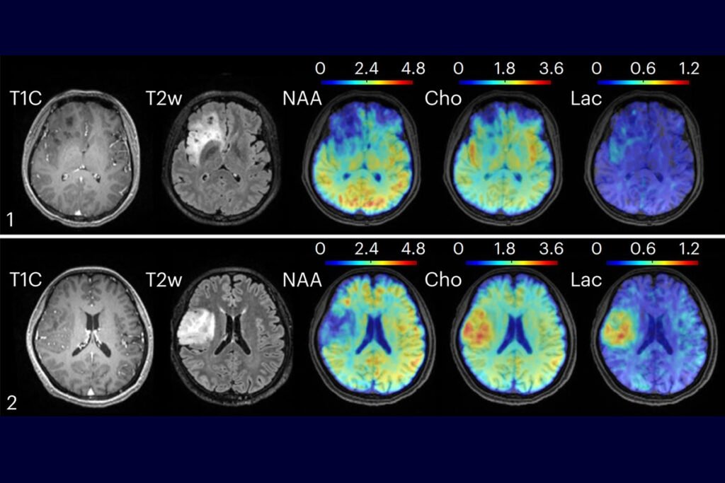
In clinical tests, the new method distinguished between healthy brain regions, identified metabolic differences in brain tumors, and even mapped lesions in multiple sclerosis (MS) patients—often before structural changes appeared on traditional MRI. For example, in patients with oligodendroglioma, a type of brain tumor, the system detected elevated levels of choline and lactate in higher-grade tumors that looked identical on standard scans. That kind of biochemical insight could help clinicians grade tumors more accurately, monitor treatment response, and personalize care.
The technology also holds promise for neurodegenerative and inflammatory conditions. Because metabolic changes often precede structural damage, this method could catch diseases like Alzheimer’s, Parkinson’s, or MS earlier—when interventions are more likely to succeed. It could also help differentiate between similar-looking lesions or track disease progression over time, offering a more dynamic view of brain health.
What makes this breakthrough especially compelling is its accessibility. The technique runs on existing clinical MRI machines, meaning hospitals don’t need to invest in new hardware. And because the scan takes just 12 minutes, it’s practical for routine use—unlike older MRSI methods that required long scan times and produced noisy, low-resolution images. By integrating smart algorithms with fast imaging protocols, the Illinois team has effectively removed the two biggest barriers to metabolic MRI: time and clarity.
Professor Liang sees this as a natural evolution of MRI’s role in neuroscience. “Our new technology adds another dimension to MRI’s capability for brain imaging,” he said. “Visualization of brain metabolism and detection of metabolic alterations associated with brain diseases.” It’s a shift from seeing the brain as a static structure to understanding it as a living, chemical ecosystem—one that can now be mapped, monitored, and, hopefully, healed.
Article from University of Illinois: New MRI approach maps brain metabolism, revealing disease signatures
Abstract from Nature Biomedical Engineering: Ultrafast J-resolved magnetic resonance spectroscopic imaging for high-resolution metabolic brain imaging